
Keratoconus in Austin, TX
Keratoconus is a progressive corneal condition in which the normally round, dome-shaped cornea gradually thins and bulges outward into a cone-like shape. This irregular curvature distorts the way light enters the eye, causing increasingly blurry, distorted, and glare-sensitive vision that standard glasses or soft contact lenses cannot fully correct as the condition advances. At Freedom Eye Care in Austin, TX, we diagnose and monitor keratoconus using corneal topography and OCT imaging, and we provide specialized contact lens fitting to help patients with keratoconus achieve the clearest, most comfortable vision possible.
Book OnlineSymptoms and Progression of Keratoconus
Keratoconus typically begins in the teenage years or early 20s and progresses — sometimes rapidly, sometimes slowly — over the following one to two decades before stabilizing in most patients by their 30s or 40s. Early keratoconus may present as frequent prescription changes, increasing astigmatism that does not fit a typical pattern, and mildly blurry vision that responds to glasses but worsens between updates. As the cone progresses, symptoms become more pronounced:
- Increasing blurry or distorted vision that glasses no longer adequately correct
- Monocular diplopia (seeing a “ghost” image or multiple images with one eye)
- Halos, glare, and streaking around lights — particularly at night
- Increased sensitivity to light and glare
- Eye rubbing (which is also a known risk factor for progression)
- Corneal hydrops in advanced cases — a sudden, painful clouding of the cornea caused by a rupture in Descemet’s membrane
Because keratoconus mimics high astigmatism in its early stages, it is frequently misdiagnosed or missed without the benefit of corneal topography. At Freedom Eye Care, we include topographic screening as part of our evaluation for any patient with irregular astigmatism, rapidly changing prescriptions, or other clinical features suggesting keratoconus.
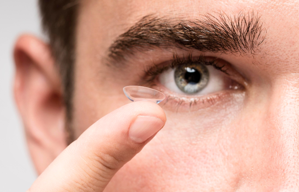
Contact Lens Management for Keratoconus
As keratoconus advances beyond what soft toric lenses can effectively correct, specialty contact lenses become the primary means of achieving functional vision. The principle behind all specialty keratoconus lenses is the same: the rigid lens creates a smooth, regular refracting surface over the irregular cone, correcting the optical distortion that the misshapen cornea would otherwise cause. Options include:
- Rigid Gas-Permeable (RGP) Lenses: The traditional specialty lens for keratoconus. RGP lenses vault over the cone and provide excellent optical correction, though some patients find the initial adaptation challenging.
- Scleral Lenses: Larger diameter lenses that vault entirely over the cornea and rest on the less sensitive sclera. Scleral lenses are the preferred option for many keratoconus patients because they provide superior comfort, stability, and vision quality compared to corneal RGP lenses — and the fluid reservoir between the lens and cornea also benefits patients with associated dry eye. Our contact lens fitting team is experienced with scleral lens fitting for keratoconus.
- Hybrid Lenses: A rigid optical center bonded to a soft skirt, offering the visual clarity of an RGP with enhanced comfort from the soft outer zone.
- Piggyback Systems: Wearing a soft lens under an RGP to improve comfort for patients who cannot tolerate RGP lenses directly on the cornea.
For patients with progressive keratoconus, corneal cross-linking — a procedure that strengthens the corneal tissue to halt progression — is performed by corneal specialists and has become the standard of care for halting keratoconus progression. Freedom Eye Care provides referrals to trusted corneal specialists in Austin, TX when cross-linking is indicated, and continues to manage your contact lens correction and eye health monitoring before and after any procedure. Call us at (512) 916-4600 or schedule your keratoconus evaluation online.
Frequently Asked Questions About Keratoconus in Austin, TX
Is keratoconus hereditary?
There is a genetic component to keratoconus — approximately 10% of patients have a family member with the condition, and the risk is higher if a first-degree relative is affected. However, most keratoconus occurs without an obvious family history. Associated conditions including Down syndrome, atopic disease (eczema, asthma, allergies), and connective tissue disorders are associated with higher keratoconus prevalence. Genetic testing is not routinely used in diagnosis; corneal topography remains the gold standard screening tool at Freedom Eye Care.
Why is eye rubbing a risk factor for keratoconus?
Frequent and forceful eye rubbing applies repeated mechanical stress to the cornea, which can damage the structural collagen fibers that give the cornea its shape and strength. In individuals who are genetically predisposed to keratoconus, this repeated trauma is believed to accelerate or trigger corneal thinning and ectasia. Patients with keratoconus — and those at risk — are strongly advised to avoid eye rubbing. Treating the underlying cause of the rubbing (typically dry eye or allergies) is often the most effective way to break the habit.
Can keratoconus be detected before symptoms appear?
Yes — and early detection is one of the most important goals of keratoconus screening at Freedom Eye Care. Corneal topography can identify subtle irregularities in corneal shape that precede the onset of noticeable symptoms by years. Identifying keratoconus at an early subclinical stage — particularly in teenagers and young adults — creates the opportunity to monitor progression and consider corneal cross-linking to halt it before significant distortion and vision loss develop.
What is corneal cross-linking and does it cure keratoconus?
Corneal cross-linking (CXL) is a minimally invasive procedure in which riboflavin (vitamin B2) eye drops are applied to the cornea and then activated with ultraviolet-A light. This creates new bonds between the collagen fibers in the corneal stroma, significantly strengthening the tissue and halting the progressive thinning and bulging of keratoconus. Cross-linking does not reverse existing corneal distortion or eliminate the need for specialty lenses — it stops further progression. Freedom Eye Care refers patients for cross-linking when indicated and co-manages care throughout the process.
Will I eventually need a corneal transplant for keratoconus?
The majority of keratoconus patients — particularly those diagnosed today, when cross-linking and specialty lenses are widely available — do not progress to the point of requiring a corneal transplant. Historically, before cross-linking became standard of care, approximately 10–20% of keratoconus patients eventually required transplantation. With timely cross-linking to halt progression and appropriate specialty lens fitting to maintain functional vision, most patients achieve excellent outcomes without surgery. Freedom Eye Care monitors your progression closely and makes referrals at the appropriate time if surgical intervention becomes necessary.
Can I wear regular soft contact lenses if I have keratoconus?
In early keratoconus, standard soft toric lenses may provide adequate vision correction. However, as the cone progresses and the corneal irregularity increases, soft lenses drape over the irregular surface and cannot compensate for the distortion — resulting in blurry, unstable vision despite an accurate prescription. At that point, specialty rigid lenses (RGP, scleral, or hybrid) become necessary to achieve functional vision. Freedom Eye Care monitors your keratoconus progression and advises you on when to transition to specialty lenses.